Home

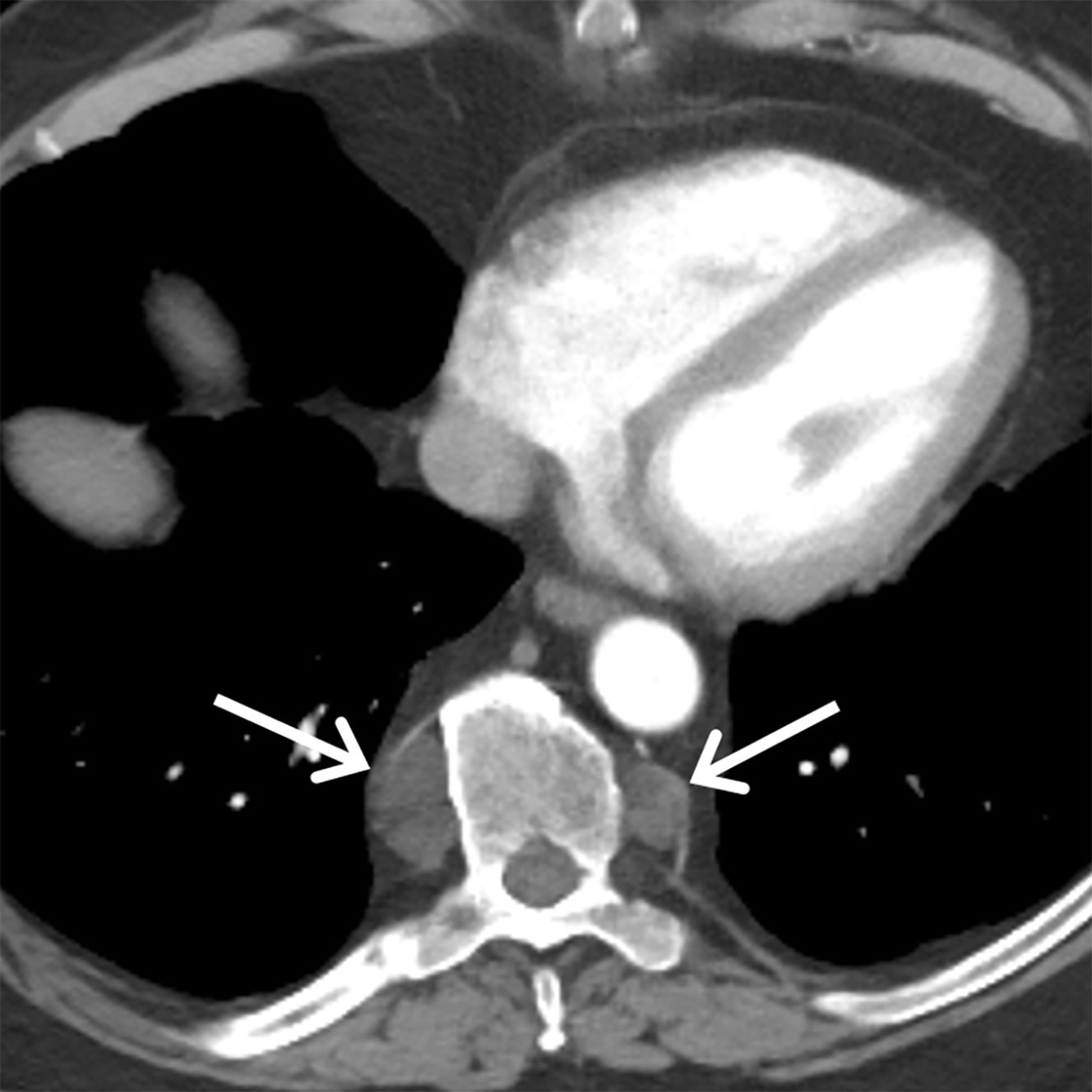
paraspinal soft tissues

Perivertebral space | Radiology Reference Article | Radiopaedia.org

Chest radiographs revealed bony destruction

MRI of the spine with paraspinal soft tissue swelling and enhancement... |  Download Scientific Diagram
MRI of the spine with paraspinal soft tissue swelling and enhancement... | Download Scientific Diagram

Imaging of the Posterior/Paravertebral Mediastinum | Radiology Key

Soft Tissue Injury - Cervical and Lumbar Strain

Chest PA radiograph shows an abnormal right paraspinal soft tissue... |  Download Scientific Diagram
Chest PA radiograph shows an abnormal right paraspinal soft tissue... | Download Scientific Diagram

Paravertebral soft tissue mass (a) before and (b) after treatment with... |  Download Scientific Diagram
Paravertebral soft tissue mass (a) before and (b) after treatment with... | Download Scientific Diagram

Understanding a mass in the paraspinal region: an anatomical approach |  Insights into Imaging | Full Text
Understanding a mass in the paraspinal region: an anatomical approach | Insights into Imaging | Full Text

2.4 Paravertebral soft tissue mass with displacement of the left... |  Download Scientific Diagram
2.4 Paravertebral soft tissue mass with displacement of the left... | Download Scientific Diagram

Fig 3. | Complex Spinal-Paraspinal Fast-Flow Lesions in CLOVES Syndrome:  Analysis of Clinical and Imaging Findings in 6 Patients | American Journal  of Neuroradiology
Fig 3. | Complex Spinal-Paraspinal Fast-Flow Lesions in CLOVES Syndrome: Analysis of Clinical and Imaging Findings in 6 Patients | American Journal of Neuroradiology

Paraspinal lesions consistent with extra-medullary hematopoiesis. images,  diagnosis, treatment options, answer review - Thoracic Imaging
Paraspinal lesions consistent with extra-medullary hematopoiesis. images, diagnosis, treatment options, answer review - Thoracic Imaging

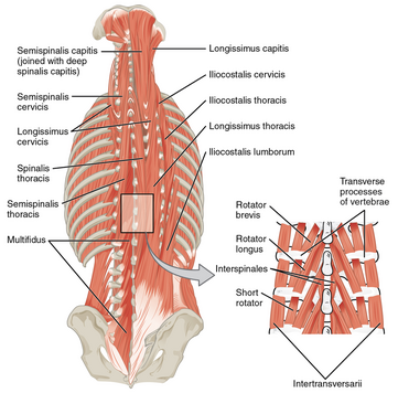
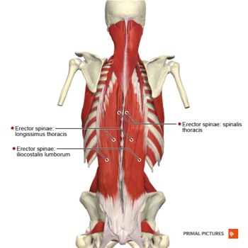
Paraspinal Muscles - Physiopedia

Paraspinal Muscles - Physiopedia

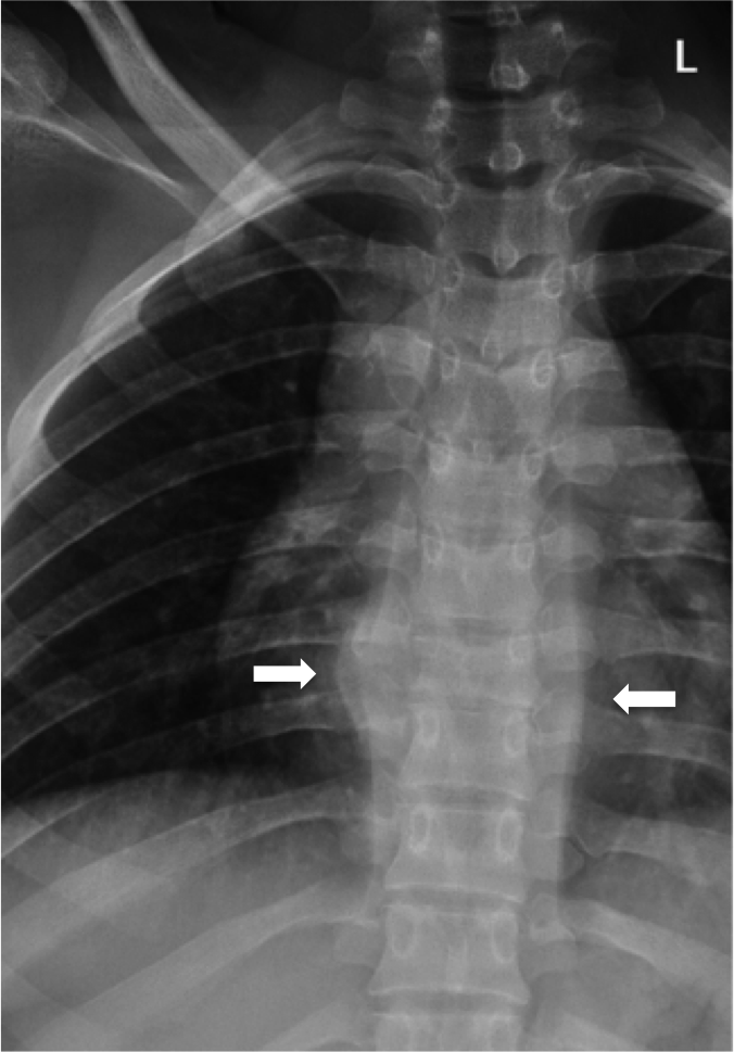
Differentiating Normal from Abnormal Inferior Thoracic Paravertebral Soft  Tissues on Chest Radiography in Children | AJR
Differentiating Normal from Abnormal Inferior Thoracic Paravertebral Soft Tissues on Chest Radiography in Children | AJR

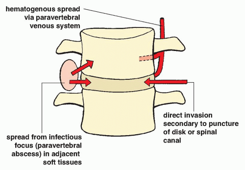
Radiologic Evaluation of Musculoskeletal Infections | Radiology Key

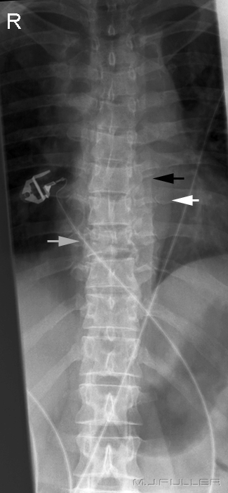
Soft Tissue Signs- Thoracic Spine - wikiRadiography

Lumbar Strain - Physiopedia

Acute paraspinal compartment syndrome following non-spinal orthopedic  surgery
Acute paraspinal compartment syndrome following non-spinal orthopedic surgery

EPOS™
EPOS™

Understanding a mass in the paraspinal region: an anatomical approach |  Insights into Imaging | Full Text
Understanding a mass in the paraspinal region: an anatomical approach | Insights into Imaging | Full Text

A) 59-year-old woman, paravertebral soft tissue type. (A, B and C)... |  Download Scientific Diagram
A) 59-year-old woman, paravertebral soft tissue type. (A, B and C)... | Download Scientific Diagram

Understanding a mass in the paraspinal region: an anatomical approach |  Insights into Imaging | Full Text
Understanding a mass in the paraspinal region: an anatomical approach | Insights into Imaging | Full Text

Reconstruction of Spinal Soft Tissue Defects With Perforator Flaps From the  Paraspinal Region | In Vivo
Reconstruction of Spinal Soft Tissue Defects With Perforator Flaps From the Paraspinal Region | In Vivo

EPOS™
EPOS™

Differentiating Normal from Abnormal Inferior Thoracic Paravertebral Soft  Tissues on Chest Radiography in Children | AJR
Differentiating Normal from Abnormal Inferior Thoracic Paravertebral Soft Tissues on Chest Radiography in Children | AJR

Understanding a mass in the paraspinal region: an anatomical approach |  Insights into Imaging | Full Text
Understanding a mass in the paraspinal region: an anatomical approach | Insights into Imaging | Full Text

Understanding a mass in the paraspinal region: an anatomical approach |  Insights into Imaging | Full Text
Understanding a mass in the paraspinal region: an anatomical approach | Insights into Imaging | Full Text

New Grading System for Cervical Paraspinal Soft Tissue Damage After  Traumatic Cervical Spinal Cord Injury Without Major Fracture Based on the  Short-T1 Inversion Recovery Mid-Sagittal MRI for Prediction of Neurological  Improvements: The
New Grading System for Cervical Paraspinal Soft Tissue Damage After Traumatic Cervical Spinal Cord Injury Without Major Fracture Based on the Short-T1 Inversion Recovery Mid-Sagittal MRI for Prediction of Neurological Improvements: The